The seed sequence or seed region is a conserved heptametrical sequence which is mostly situated at positions 27 from the miRNA 5´end Even though base pairing of miRNA and its target mRNA does not match perfect, the "seed sequence" has to be perfectly complementaryThe fine brain region in the rough brain mask is segmented using multiseeded region growing approach The proposed method uses multiple seed points which are selected automatically based on the intensity profile of grey matter (GM), white matter (WM) and cerebrospinal fluid (CSF) of the brain1521 · Tellus Therapeutics, Inc today announced a seed investment from Xontogeny, LLC to advance their lead program through INDenabling work

Whole Brain Differences In Mtl Functional Connectivity Seed Regions Download Scientific Diagram
Is there a seed in every brain
Is there a seed in every brain-Seedbased connectivity metrics characterize the connectivity patterns with a predefined seed or ROI (Region of Interest) These metrics are often used when researchers are interested in one, or a few, individual regions and would like to analyze in detail the connectivity patterns between these areas and the rest of the brain · Seedbased functional connectivity, also called ROIbased functional connectivity, finds regions correlated with the activity in a seed region In seedbased analysis, the crosscorrelation is computed between the timeseries of the seed and the rest of the brain (telling us where the traffic is communicating between selected cities) (Fig 3, the results are visualized with


Mapping Cortical Network Effects Of Fatigue During A Handgrip Task By Functional Near Infrared Spectroscopy In Physically Active And Inactive Subjects
(ii) moderately increased striatal correlations with specific parts of the cerebral cortex; · Taskrelated brain activity was detected under saline in areas involved in memory, pain, and fear, particularly the hippocampus, insula, and amygdala Compared with saline, midazolam increased functional connectivity to brain areas and decreased to 8, from seed regions in the precuneus, posterior cingulate, and left insulaThe TNH group displayed increased connectivity, compared to the HC group, in brain regions that encompassed the left IC, bilateral Heschl gyrus, bilateral supplementary motor area, right insula, bilateral superior temporal gyrus, right middle temporal gyrus, left hippocampus, left amygdala, and right supramarginal gyrus
0619 · Specific connectivity with Operculum 3 (OP3) brain region in acoustic trauma tinnitus a seedbased resting state fMRI study View ORCID Profile Agnès Job , View ORCID Profile Anne Kavounoudias , Chloé Jaroszynski , Assia Jaillard , View ORCID Profile Chantal DelonMartinThe brain image (Gonzalez and Wood s, 08, Vishnuvarthanan et al, 16) The proposed Region Growing methodology helps in effectively identifying the tumor part and eas es the complication in identifying the tumor infiltrated region done manually by a radiologist who has acquaintance with radio surgery applicationsThis tumor looks just like ovarian or testicular cancer under the microscope It is the most common of the germ cell tumors of the brain It may spread or "seed" through the spinal fluid About one third of tumors in the pineal region are germinomas;
2416 · How to find seed point for region growing Learn more about brain tumor segmentation, image segmentation, region growing Image Processing Toolbox4) Brain tumor detection and area calculation The following methods achieve the brain tumor segmentation from the brain MRIs Initially, the input brain MRIs are preprocessed and converted into a binary image by thresholding technique and morphological operation The thresholding =Simple but effective example of "Region Growing" from a single seed pointThe region is iteratively grown by comparing all unallocated neighbouring pixels to


Anatomical And Functional Organization Of The Human Substantia Nigra And Its Connections Elife
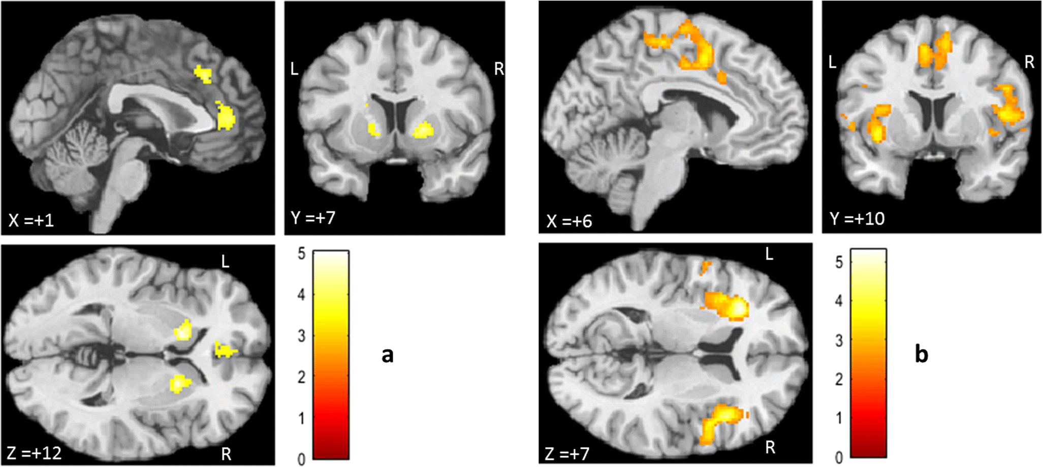


Figure 2 Aberrant Functional Connectivity Profiles Of Brain Regions Associated With Salience And Reward Processing In Female Patients With Borderline Personality Disorder Springerlink
Of brain lesions from diffusionweighted magnetic resonance imaging (DWMRI or DWI) using region growing approach The lesions are acute infarction, haemorrhage, tumour and abscess Region splitting and merging is used to detect the lesion region Then, histogram thresholding technique is applied to automate the seeds selection The region is · Greater brain resting state connectivity in fatigued BC survivors compared to nonfatigued survivors Brain images show greater intrinsic connectivity between seed (left) to other brain regions (middle) Bar graphs (right) show the level of connectivity between seed and connected region in both groups using Fisher transformed Rvalues (yaxis) · Westhoff, A Hybrid parallelization of a seeded region growing segmentation of brain images for a GPU cluster In ARCS 14 27th International Conference on Architecture of Computing Systems Workshop Proceedings, p 8 Berlin, Lübeck (Germany), 25 Feb 14–28 Feb 14, VDE Verlag, February 14 Google Scholar


Sustained Anxiety Increases Amygdala Dorsomedial Prefrontal Coupling A Mechanism For Maintaining An Anxious State In Healthy Adults Abstract Europe Pmc


Brain Network Organization During Mindful Acceptance Of Emotions Biorxiv
Region growing (also called region merging) is a technique for extracting a connected region of the image which consists of groups of pixels/voxels with similar intensities In its simplest form, region growing starts with a seed point (pixel/voxel) that belongs to the object of interest2705 · Seedbased CPM analyses revealed networks that predicted higher and lower feelings of stress in novel individuals (Fig 2b–g;Thus, it displays brain regions that are coactivated across the restingstate fMRI time series with the seed voxel Values are pearson correlations (r) To reduce blurring of signals across cerebrocerebellar and cerebrostriatal boundaries, fMRI signals from adjacent cerebral cortex are regressed from the cerebellum and striatum


Resting State Functional Mri Everything That Nonexperts Have Always Wanted To Know American Journal Of Neuroradiology


Resting State Functional Mri Everything That Nonexperts Have Always Wanted To Know American Journal Of Neuroradiology
Seedbased Correlation Analysis (SCA) is one of the most common ways to explore functional connectivity within the brain Based on the time series of a seed voxel (or ROI), connectivity is calculated as the correlation of time series for all other voxels in the brain The result of SCA is a connectivity map showing Zscores for each voxelSegmentation of brain MRI in an image sequence is one of the most challenging problems in image processing, while at the same time one that finds numerous applications In this paper, we propose a robust multilayer background subtraction technique and seed region growing approach which takes advantages of local texture features represented by local binary patterns (LBP) and · Using seed regions of interest manually defined bilaterally in three subdivisions of the striatum, the principal findings in Parkinson's disease were as follows (i) markedly lower striatal correlations with the extended brainstem (ie thalamus, midbrain, upper pons and cerebellum);


A Connectome Wide Functional Signature Of Trait Anger Biorxiv


Plos One The Effect Of Criticism On Functional Brain Connectivity And Associations With Neuroticism
Automated tumor segmentation in MR brain image using fuzzy cmeans clustering and seeded region methodology Mustafa Zuhaer Nayef ALDabagh Department of Computer Science, · Automatic Seeded Selection Region Growing Algorithm for Effective MRI Brain Image Segmentation and ClassificationProbabilistic vs deterministic fiber tracking and the influence of different seed regions to delineate cerebellarthalamic fibers in deep brain stimulation Eur J Neurosci 17 Jun;45(12) doi /ejn


Resting State Fmri Wikipedia


Mapping Cortical Network Effects Of Fatigue During A Handgrip Task By Functional Near Infrared Spectroscopy In Physically Active And Inactive Subjects
· This also means that we can place a seed voxel or seed region into one location of the brain, and it will be roughly in the same spot for every subject in our analysis To do this, we will focus on one of the core components of the socalled "restingstate network," which is a reliable pattern of connectivity observed when subjects are at restSeeded Region Growing is an integrated method brought up by Adams and Bischof 413, in which few initial seeds are generated, and more similar neighboring regions are then combined to achieve region growing 1421 In addition, the method of unsupervised vector seeded region growing suitable for medical multispectral images was established · Since 09, evidence has accumulated to suggest that Tau aggregates form first in a small number of brain cells, from where they propagate to other regions, resulting in neurodegeneration and disease Propagation of Tau aggregates is often called prionlike, which refers to the capacity of an assembled protein to induce the same abnormal conformation in a


Seed To Voxel Analysis With Calcarine As The Seed The Seed Region Mni Download Scientific Diagram


Effects Of Early And Late Bilingualism On Resting State Functional Connectivity Journal Of Neuroscience
Bub AM Segmentation of brain xray CT images using seeded region growing Thesis University of Cape Town ,Faculty of Engineering & the Built Environment ,Department of Electrical Engineering, 1996 cited yyyy month ddI working on region growing algorithm implementation in python But when I run this code on output I get black image with no errors Use CV threshold function on input image and for seed value I use mouse click to store x,y values in tuple · Pokémon characters have their own peasized region in brain, study finds Stanford study scanned brains of "Pokémon experts" and compared to a control group Jennifer Ouellette May 6, 19 300


The Anatomical Location And Volume Of Each Seed Region Is Depicted Download Scientific Diagram


1
Seedbased functional connectivity analyses and independent components analysis have been used to identify brain regions that show correlated spontaneous lowfrequency BOLD signal fluctuations 37,38 This pattern of intercorrelations over time can then be attributed to discrete, functionally connected brain networksFor partially complementary microRNAs to recognise their targets, nucleotides 2–7 of the miRNA (its 'seed region') must be perfectly complementary Animal miRNAs inhibit protein translation of the target mRNA (this is present but less common in plants)Open this seed point text file in RegionsOpen region and change its region type from "ROI" to "Seed" Click "Fiber Tracking" button to start fiber tracking at these points If no seed region is assigned, DSI Studio will use the whole brain region as the seed region
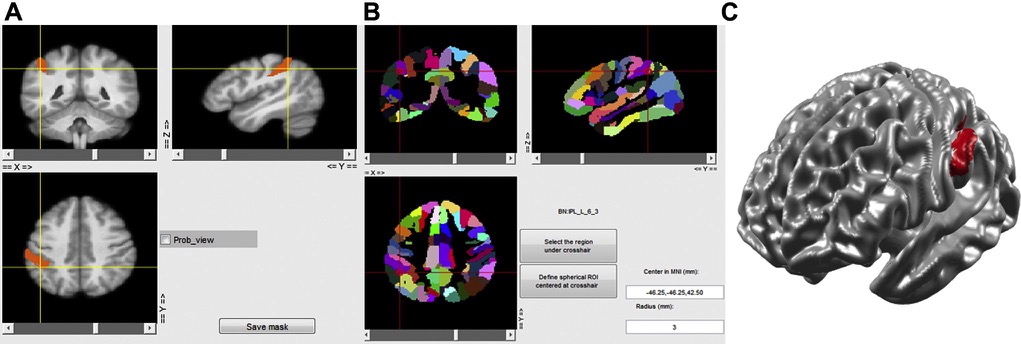


Epos Trade


Figure 4 From Rhythmic Alternating Patterns Of Brain Activity Distinguish Rapid Eye Movement Sleep From Other States Of Consciousness Semantic Scholar
Segmentation of Magnetic Resonance Imaging (MRI) images is the most challenging problems in medical imaging This paper compares the performances of SeedBased Region Growing (SBRG), Adaptive NetworkBased Fuzzy Inference System (ANFIS) and Fuzzy cMeans (FCM) in brain abnormalities segmentation Controlled experimental data is used, which designed in such a wayHowever, this tumor can occur in many locations within the brain · Group exponential lasso models were then used to predict gene cluster expression summaries as a function of seed region structural connectivity patterns In several gene clusters, brain regions located in the brain stem, diencephalon, and hippocampal formation were identified that have significant predictive power for these expression summaries


3 Dimensional Brain Mri Segmentation Based On Multi Layer Background Subtraction And Seed Region Growing Algorithm Scientific Net


Brain Connectivity And Neurological Disorders After Stroke Abstract Europe Pmc
· During performance of attentiondemanding cognitive tasks, certain regions of the brain routinely increase activity, whereas others routinely decrease activity In this study, we investigate the extent to which this taskrelated dichotomy is represented intrinsically in the resting human brain through examination of spontaneous fluctuations in the functional MRI bloodCortical thicknessdriven network analysis of the whole brain using MFC subregions as the seed region By Hyunjin Park (), YeongHun Park (), Jungho Cha (), Sang Won Seo (), Duk L Na () and JongMin Lee ()(iii) generally symmetric effects of Parkinson's


A Seed Region 4 From The Meta Analysis 12 In The Left Claustrum Download Scientific Diagram


Whole Brain Differences In Mtl Functional Connectivity Seed Regions Download Scientific Diagram
Seeded Region Growing is an integrated method brought up by Adams and Bischof 413, in which few initial seeds are generated, and more similar neighboring regions are then combined toA simple yet accurate method for segmenting magnetic resonance (MR) brain images has been implemented The semiautomatic technique carries out region growing in all three dimensions guided by initial seed points Seed voxels may be specified interactively with a mouse or automatically through the selection of intensity thresholds The 3D seeded region growing (3DRESULTS Localization of the supplementary motor area using hand motor seed regions was more effective than seeding using orofacial motor regions for both patients with brain tumor (955% versus 348%, P < 001) and controls (952% versus 452%, P < 001) Bilateral hand motor seeding was superior to unilateral hand motor seeding in patients with


Changes In Resting State Functional Brain Activity Are Associated With Waning Cognitive Functions In Hiv Infected Children Abstract Europe Pmc
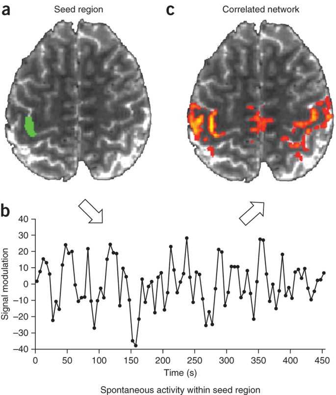


Opportunities And Limitations Of Intrinsic Functional Connectivity Mri Nature Neuroscience
Using recombinant CαSyn and PD and DLB brain lysates as seeds in the realtime quakinginduced conversion (RTQuIC) assay, we explored how CαSyn may be involved in disease stratification Comparing the seeding activity of aqueoussoluble fractions to detergentsoluble fractions, and using αSyn 1130 as substrate for the RTQuIC assay, the temporal cortex seeds · Fig 3 αSynuclein strains target distinct brain regions in TgM mice a, Semiquantitative PSyn deposition scoring within the indicated brain regions from clinically ill TgM mice followingThis paper proposes an empirical study of the efficiency of the SeedBased Region Growing (SBRG) in segmentation of brain abnormalities Presently, segmentation poses one of the most challenging problems in medical imaging Segmentation of Magnetic Resonance Imaging (MRI) images is an important part of brain imaging research


Enhanced Medial Prefrontal Default Mode Network Functional Connectivity In Chronic Pain And Its Association With Pain Rumination Journal Of Neuroscience


Tracing Connections In The Brain To Reveal Functions Kurzweil
IES mapping amplitude correlations of a seed region with the rest of the brain during restExtracting the BOLD time course from a seed region then computing the correlation coefficient between that time course and the time course from all other brain voxels Seed regions were 12mmdiameter spheres centered on previously published foci For the current study we examined correlations associated


Arxiv Org Pdf 1812


Seed Region Connectivity Map Seed Regions Are Presented On T1 Images Download Scientific Diagram


The Relationship Between Meg Networks And Myelination A Structural Download Scientific Diagram


Figure 3 From Atypical Neural Self Representation In Autism Semantic Scholar


Plos One Resting State Fmri Reveals Diminished Functional Connectivity In A Mouse Model Of Amyloidosis


Salience Network Wikipedia


1


Region Specific Protein Misfolding Cyclic Amplification Reproduces Brain Tropism Of Prion Strains Journal Of Biological Chemistry
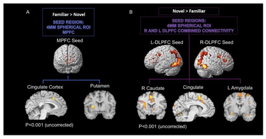


Brain Sciences Free Full Text Common And Distinct Functional Brain Networks For Intuitive And Deliberate Decision Making Html


Resting State Functional Mri Everything That Nonexperts Have Always Wanted To Know American Journal Of Neuroradiology


Defining The Seed Region Of Interest Ppi Analysis Investigates Download Scientific Diagram


Altered Extended Locus Coeruleus In Boys With Autism Ndt


Rhythmic Alternating Patterns Of Brain Activity Distinguish Rapid Eye Movement Sleep From Other States Of Consciousness Pnas


1
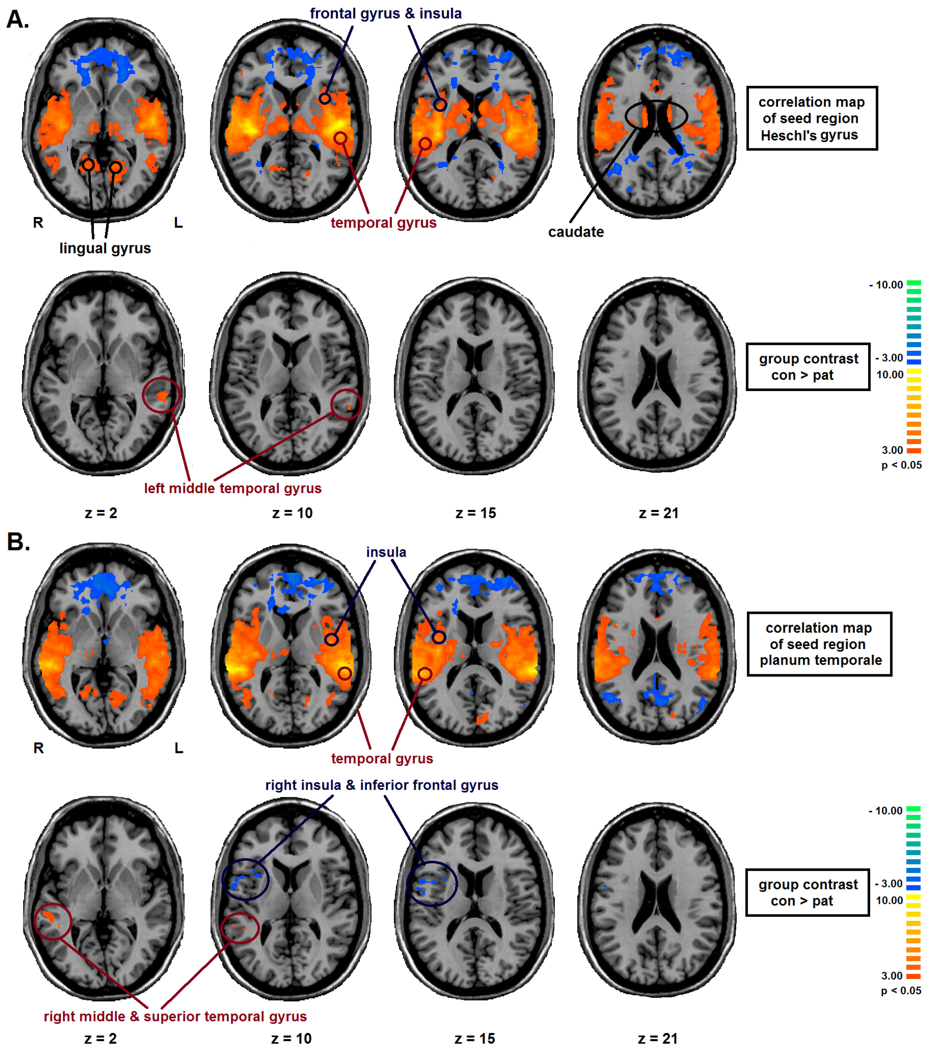


Brain Sciences Free Full Text Altered Intrinsic Functional Connectivity In Language Related Brain Regions In Association With Verbal Memory Performance In Euthymic Bipolar Patients Html


Intrinsic Brain Connectivity After Partial Sleep Deprivation In Young And Older Adults Results From The Stockholm Sleepy Brain Study Biorxiv


Location Of The Seed Regions Seeds Were Drawn From An Earlier Download Scientific Diagram


2 Ppi Connectivity Analysis Results For Left Hemispheric Cortical Seed Download Scientific Diagram


A Window Into The Brain Advances In Psychiatric Fmri
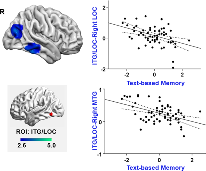


Distinct Individual Differences In Default Mode Network Connectivity Relate To Off Task Thought And Text Memory During Reading Scientific Reports


Sulcal Pattern Variability And Dorsal Anterior Cingulate Cortex Functional Connectivity Across Adult Age Brain Connectivity


1 A Location Of The Seed Region Brown And The Three Exemplary Download Scientific Diagram


Plos One Enhanced Functional Connectivity And Volume Between Cognitive And Reward Centers Of Naive Rodent Brain Produced By Pro Dopaminergic Agent Kb2z


Functional Connectivity Maps N 12 Of The Brain Using The Dn As Seed Download Scientific Diagram
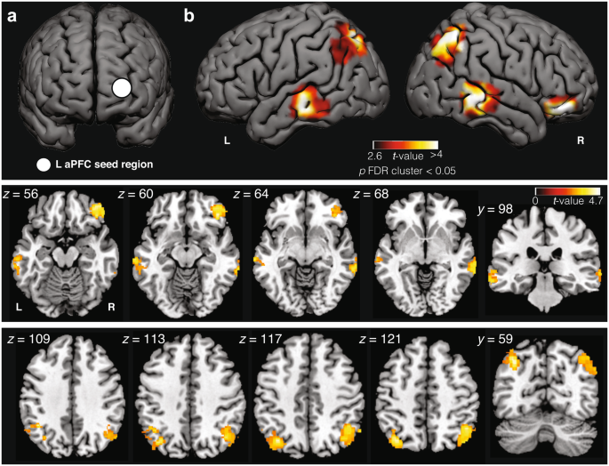


Frequent Lucid Dreaming Associated With Increased Functional Connectivity Between Frontopolar Cortex And Temporoparietal Association Areas Scientific Reports
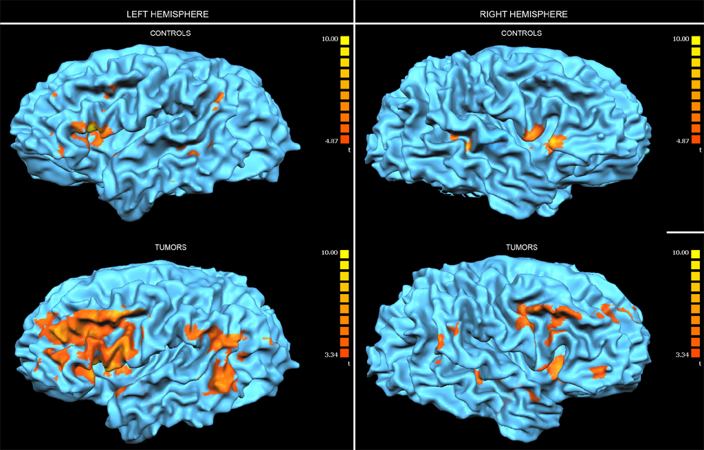


Epos Trade


Spmcourse October 13 Dcm Dynamic Causal Modelling For
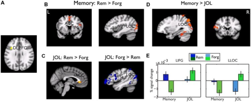


Psychophysiological Interaction Ppi Results A The Open I


Seed To Voxel Analysis With Calcarine As The Seed The Seed Region Mni Download Scientific Diagram


Http Biorxiv Org Cgi Reprint 10 29 v1


Brain Areas Showing Functional Connectivity With The Seed Region Of The Download Scientific Diagram


Natural Connection Patterns Drive Brain Development Brainpost Easy To Read Summaries Of The Latest Neuroscience Publications


1


Whole Brain Fetal Age Regression Analysis For The Pcc Seed Region Download Scientific Diagram


Figure 4 From Eeg Alpha Power Modulation Of Fmri Resting State Connectivity Semantic Scholar


Altered Hypothalamic Functional Connectivity In Cluster Headache A Longitudinal Resting State Functional Mri Study Journal Of Neurology Neurosurgery Psychiatry


Academic Oup Com Cercor Article Pdf 25 9 3046 Bhu100 Pdf


View Image
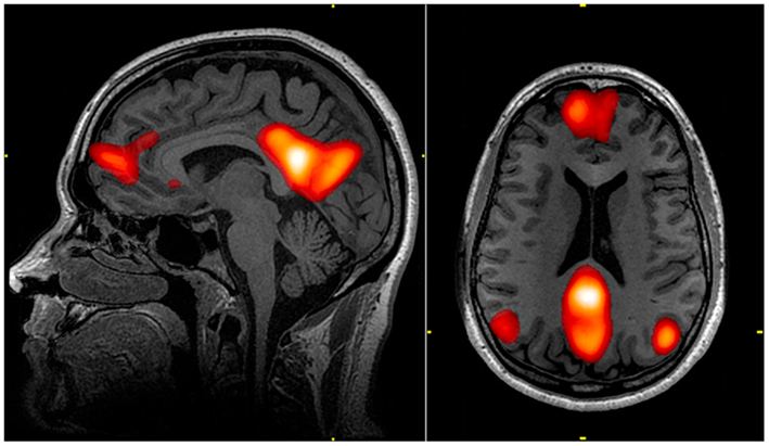


Default Mode Network Wikipedia


Figure 2 From Resting State Connectivity Immediately Following Learning Correlates With Subsequent Sleep Dependent Enhancement Of Motor Task Performance Semantic Scholar


Plos One Personality Is Reflected In The Brain S Intrinsic Functional Architecture


Brain Areas Showing Greater Functional Connectivity With The Bilateral Download Scientific Diagram


Seed Regions Of Interest Note The Salience Network Defined Using A Download Scientific Diagram


Figure 2 From Multi Echo Fmri Resting State Connectivity And High Psychometric Schizotypy Semantic Scholar


Whole Brain Connectivity Of Left Hippocampus Seed Region Left Hf And Download Scientific Diagram
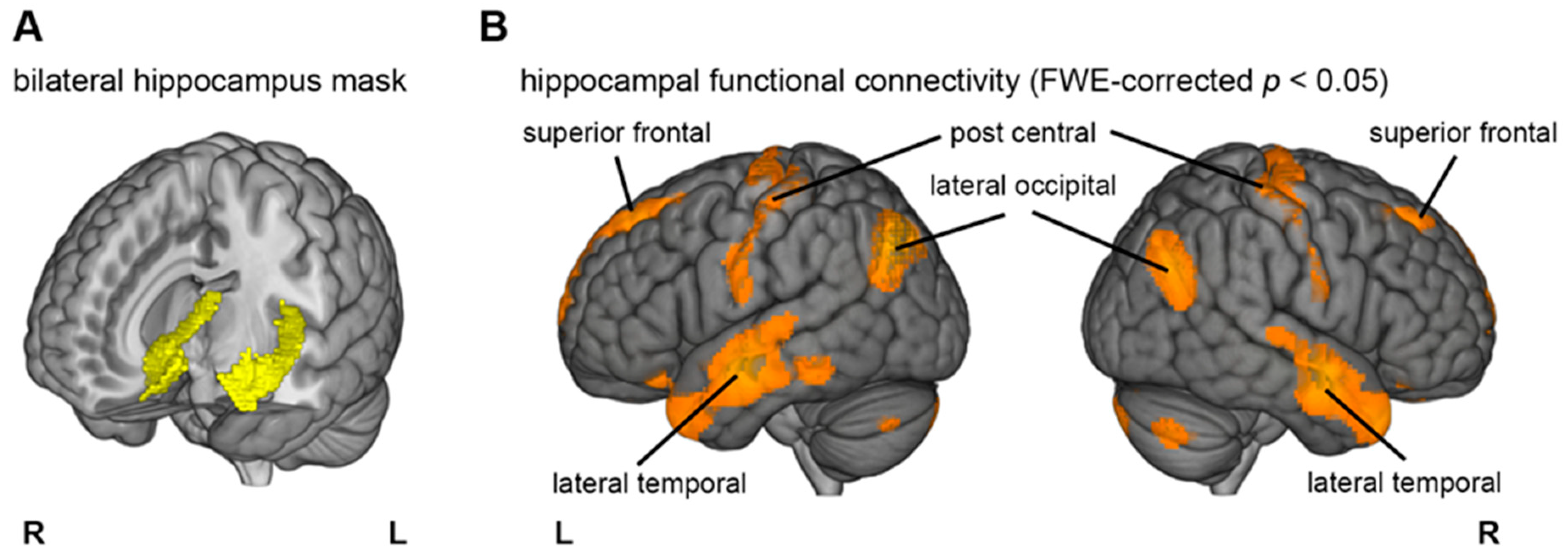


Brain Sciences Free Full Text Bi Temporal Anodal Transcranial Direct Current Stimulation During Slow Wave Sleep Boosts Slow Wave Density But Not Memory Consolidation Html


Mapping Cortical Network Effects Of Fatigue During A Handgrip Task By Functional Near Infrared Spectroscopy In Physically Active And Inactive Subjects


Right Supramarginal Gyrus Is Crucial To Overcome Emotional Egocentricity Bias In Social Judgments Abstract Europe Pmc


Spmcourse October 13 Dcm Dynamic Causal Modelling For


The Identified Brain Region Is Seed Region Where Reho Correlated Download Scientific Diagram
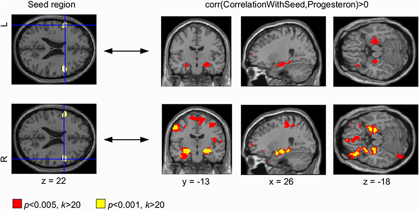


Frontiers Progesterone Mediates Brain Functional Connectivity Changes During The Menstrual Cycle A Pilot Resting State Mri Study Neuroscience


Correlation Between Traits Of Emotion Based Impulsivity And Intrinsic Default Mode Network Activity


Whole Brain Resting State Functional Connectivity Maps With Seed Region Download Scientific Diagram


The Anatomical Localization Of The Four Seed Region Of Interest Download Scientific Diagram


Seed And Target Regions The Brain Explorer Of Aba Can Represents Download Scientific Diagram
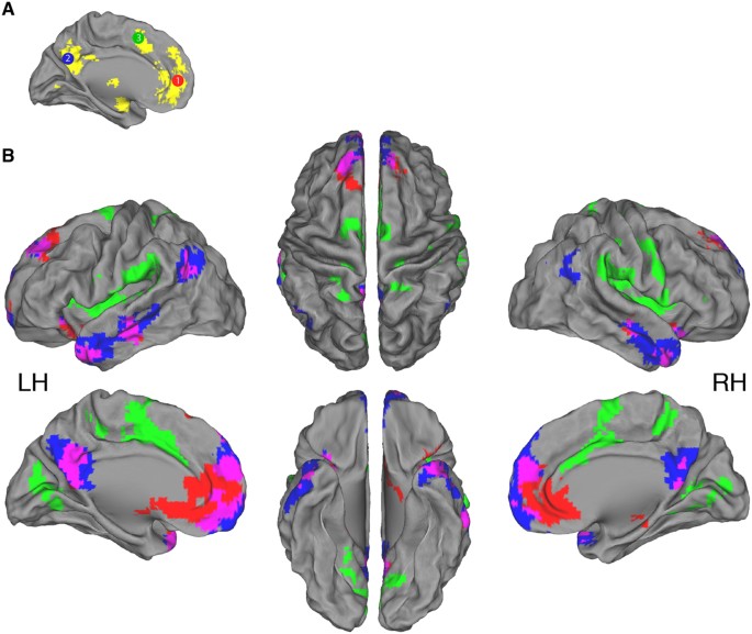


Sharing Self Related Information Is Associated With Intrinsic Functional Connectivity Of Cortical Midline Brain Regions Scientific Reports


Enhanced Medial Prefrontal Default Mode Network Functional Connectivity In Chronic Pain And Its Association With Pain Rumination Journal Of Neuroscience
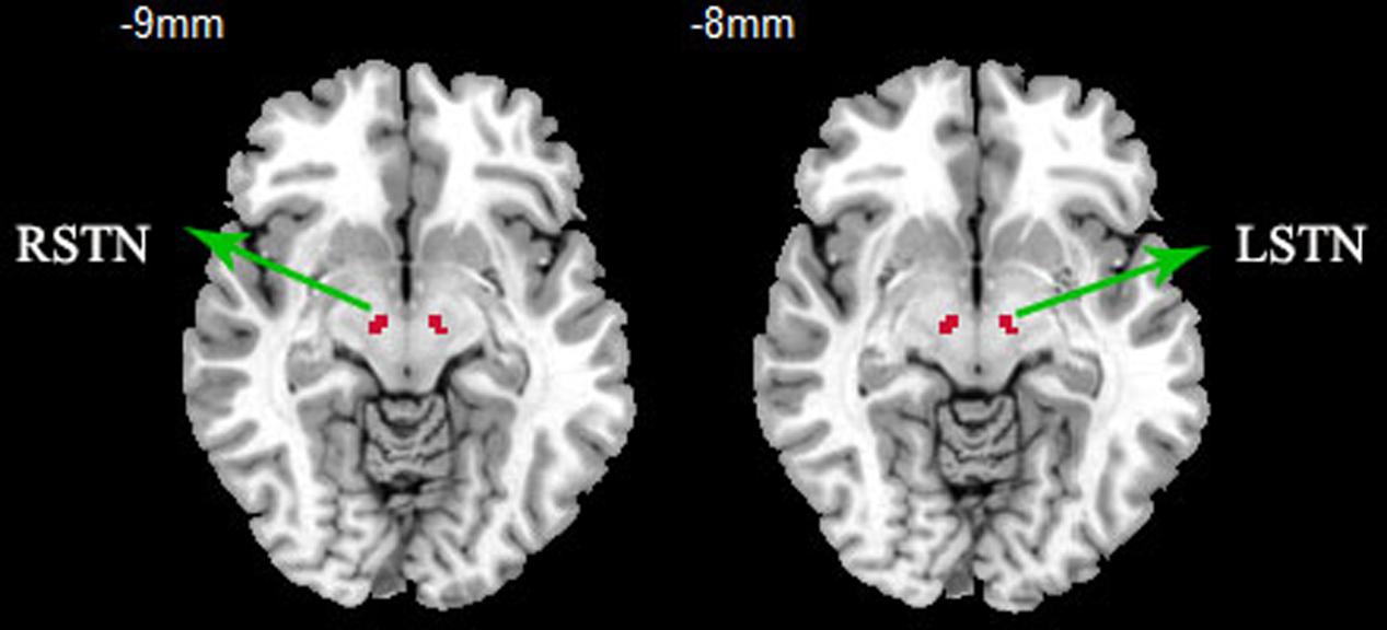


Frontiers Resting State Fmri Reveals Increased Subthalamic Nucleus And Sensorimotor Cortex Connectivity In Patients With Parkinson S Disease Under Medication Frontiers In Aging Neuroscience
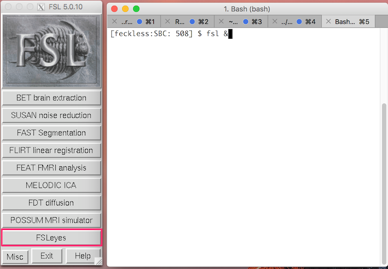


Fsl Fmri Resting State Seed Based Connectivity Neuroimaging Core 0 1 1 Documentation
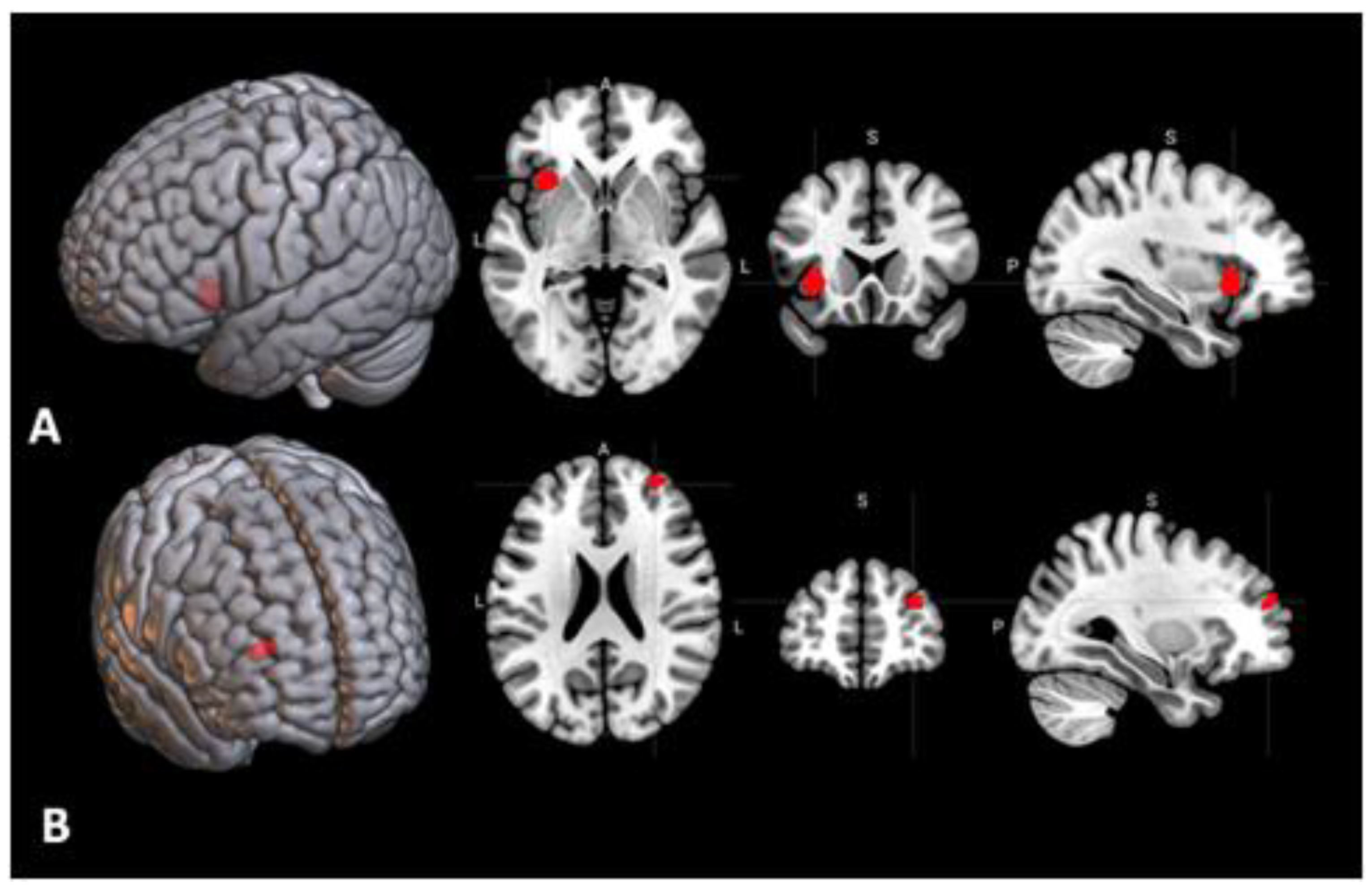


Behavioral Sciences Free Full Text Brain Activations And Functional Connectivity Patterns Associated With Insight Based And Analytical Anagram Solving Html


Task Dependent Organization Of Brain Regions Active During Rest Pnas


Illustration Of Parcellation Routine For Seed Region Node Definition Download Scientific Diagram


Figure 6 From The Impact Of Global Signal Regression On Resting State Correlations Are Anti Correlated Networks Introduced Semantic Scholar


Seizure Frequency Can Alter Brain Connectivity Evidence From Resting State Fmri American Journal Of Neuroradiology
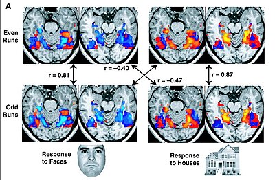


Resting State Fmri Wikipedia


Plos One Interactions Between Affective And Cognitive Processing Systems In Problematic Gamblers A Functional Connectivity Study


The Human Brain Is Intrinsically Organized Into Dynamic Anticorrelated Functional Networks Pnas


Cortical Functional Connectivity Evident After Birth And Behavioral Inhibition At Age 2 American Journal Of Psychiatry


Meditation Experience Is Associated With Differences In Default Mode Network Activity And Connectivity Pnas


Plos One Restoring Susceptibility Induced Mri Signal Loss In Rat Brain At 9 4 T A Step Towards Whole Brain Functional Connectivity Imaging


Fiber Connections From Functional Activation Seeds Tractograms For Two Download Scientific Diagram
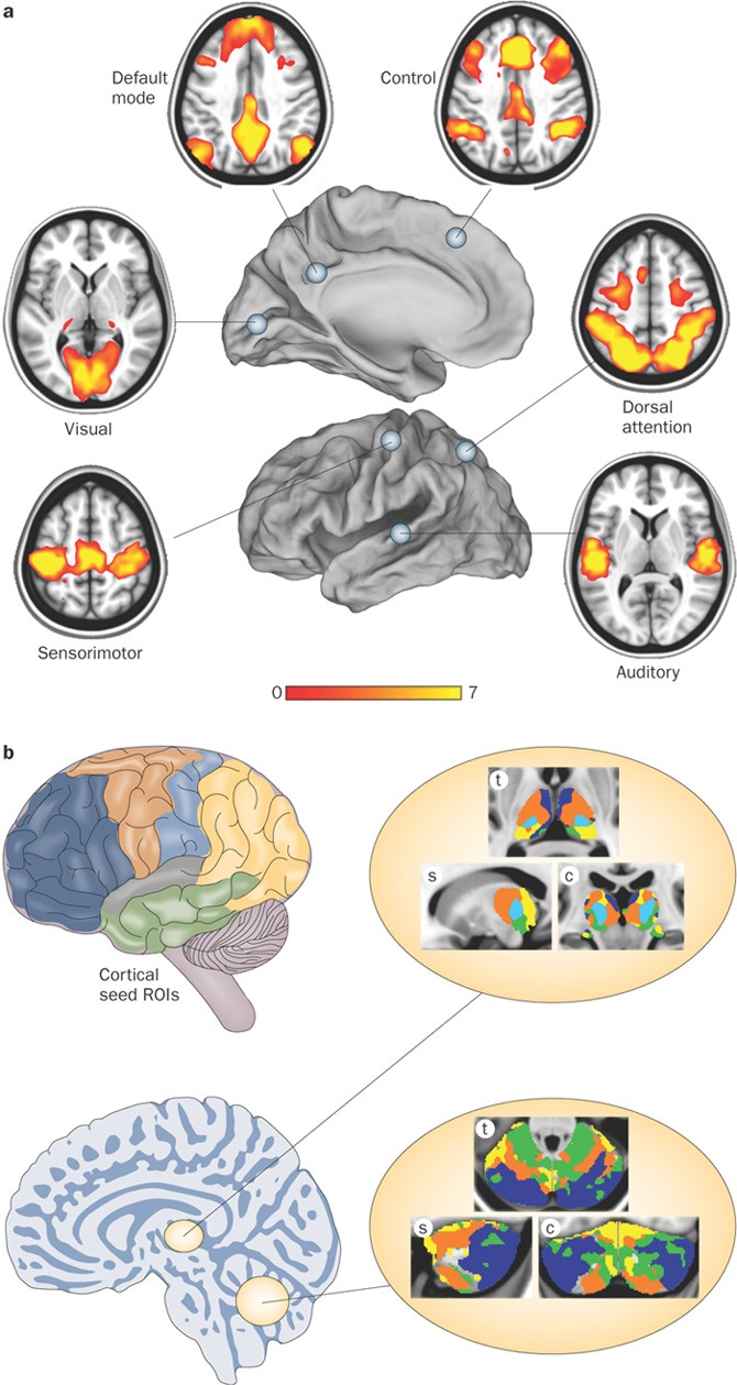


Disease And The Brain S Dark Energy Nature Reviews Neurology
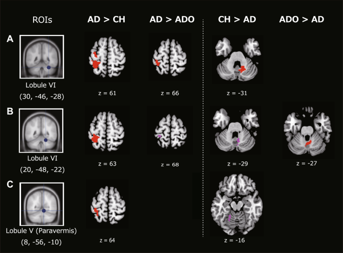


Local To Distant Development Of The Cerebrocerebellar Sensorimotor Network In The Typically Developing Human Brain A Functional And Diffusion Mri Study Springerlink


Neuroscientists Just Launched An Atlas Of The Developing Human Brain Wired


Resting State Fmri An Overview Sciencedirect Topics


Resting State Functional Mri Everything That Nonexperts Have Always Wanted To Know American Journal Of Neuroradiology


Seed Regions Of The Seed Based Resting State Analysis Seed Regions And Download Scientific Diagram


Anatomical And Functional Organization Of The Human Substantia Nigra And Its Connections Biorxiv


Resting State Functional Mri Everything That Nonexperts Have Always Wanted To Know American Journal Of Neuroradiology


Intrinsic Correlations Between A Seed Region In The Pcc And All Other Download Scientific Diagram


0 件のコメント:
コメントを投稿